Ancillary findings in distinguishing between anterior cruciate ligament mucoid degeneration and sprain on MRI: a practical approach
Sprain and mucoid degeneration are the most important and frequent pathologies of the ACL. In some cases, their distinction might be difficult both in imaging and in clinical practice
According to study of Cilengir et al. which was published in an Acta Radiologica, to overcome this difficulty;
• In cases with ACL mucoid degeneration, mean age and the female prevalence are higher, and intraosseous cyst, subchondral sclerosis, femoral and tibial chondromalacia are more frequent.
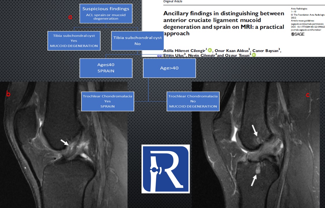
• decision tree algorithm (figure a), which provides a practical approach in the presence of suspicious finding in the ACL, achieved a total success of 79.2% by looking at only 3 data.
Figure legends: a) Decision Tree Algorithm. b) Anterior cruciate ligament partial tear in a 24-year-old male patient. Fat-suppressed proton density sagittal plane image reveals foci of fluid-like signals and partial discontinuity in the fibers of the hyperintense anterior cruciate ligament (arrow).c) Intraosseous cysts in a 54-year-old female patient. On the fat-suppressed proton density sagittal image of a patient with anterior cruciate ligament mucoid degeneration, intraosseous cyst formation (arrows) is observed on both the femoral and tibial faces
You can reach the article on;
https://journals.sagepub.com/doi/abs/10.1177/02841851221090623
Doi
https://doi.org/10.1177/02841851221090623

0 COMMENTS
These issues are no comments yet. Write the first comment...