Soft Tissue tumor
Intramuscular Lipoma
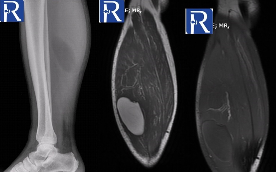
Demographic and clinical details: 53-years-old male patient, admitted with soft tissue mass in right Calf. .
Image Details: Lateral X-Ray shows the well-defined radiolucent lesion in posterior compartment. Coronal Plane T1W image shows well defined soft tissue mass that has isointense signal with fat tissue. It is seen that lesion signal is completely suppressed in fat saturated T1W image. Radiological findings is consistent with soft tissue lipoma. Radiography may give some clues l for the diagnosis of soft tissue lesion. Note the lesion density is lower that muscle of cyst but higher than air, which may suggest the fat density.

0 COMMENTS
These issues are no comments yet. Write the first comment...