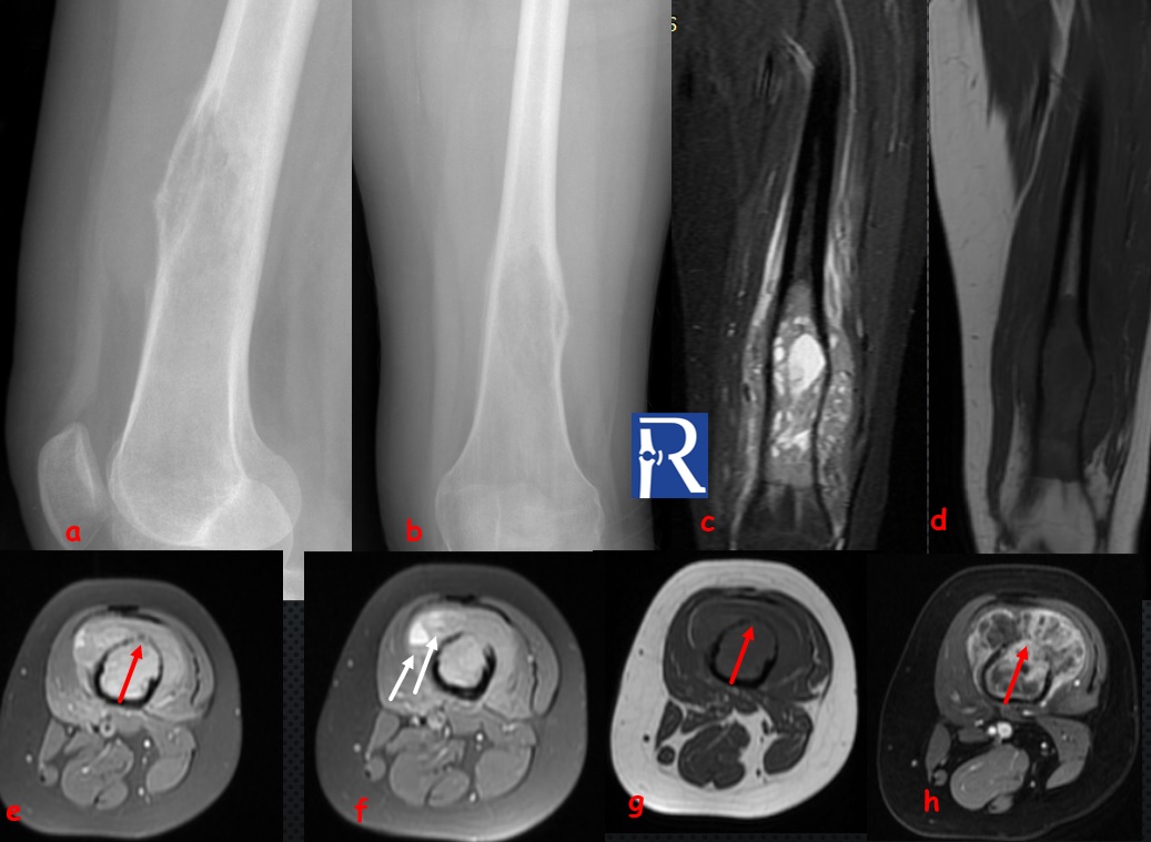
Telangiectatic osteosarcoma

Demographic and clinical details: 17 years old female, with a left thigh pain
Image Details: 2 view X-Ray of left femur (a, b) show the type 1 c geographic lytic lesion located in distal diaphysis of left femur, with expansion and thinning of anterior cortex. Coronal STIR © and T1W (d), Axial PD Fat saturated (e, f) T1 W(g) images shows the extension of the heterogenous mass lesion into the soft tissues of anterior compartment. Note fluid levels (white arrows) of small cystic cavities (white arrows) and thinning of anterior cortex with permeation regions (red arrows). Contrast enhanced axial T1W (h) fat saturated image show enhancing solid components and multiple thick septa. Radiographic findings consistent with aggressive lesion. MRI features should suggest firstly the diagnosis of telangiectatic osteosarcoma (Tel-OS.) but diagnosis of secondary aneurysmal bone cyst should be ruled out. Biopsy confirmed the diagnosis of Tel-OS
Tel-OS is a rare variant variant of OS (%1.2-7 of all osteosarcomas) which considered a subtype of osteosarcoma (not othervise specified).Tumors are consist of large blood-filled spaces separated by thin bony septations. Osteodi matrix mineralisation is less than conventional OS. Tel-OS’s are associated with high rate of pathological fracture. It should be differentiated from aneurysmal bone cyst (ABC).
In my experience, MRI shows the fluid-fluid levels in most of the cases. It should be differentiated with ABC. Tel-OS should be excluded firstly in cases with lesions that show fluid-fluid level and have thick enhancing septa and solid component..


0 COMMENTS
These issues are no comments yet. Write the first comment...