Decoding the Operated Shoulder — Sagittal Plane Secrets (Latarjet Case)
Case Presentation
Patient Information:
A 33-year-old male patient presented with persistent shoulder pain two months after undergoing surgery at an outside institution for recurrent dislocations of the left shoulder.
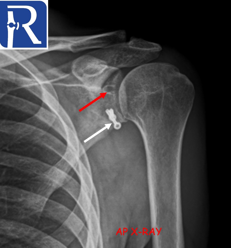
Plain Radiography Findings:
Metallic anchor-related radiopacities are seen at the inferior glenoid (white arrows).
At the superior glenoid, a radiolucent area consistent with a bioabsorbable anchor is observed (red arrows).
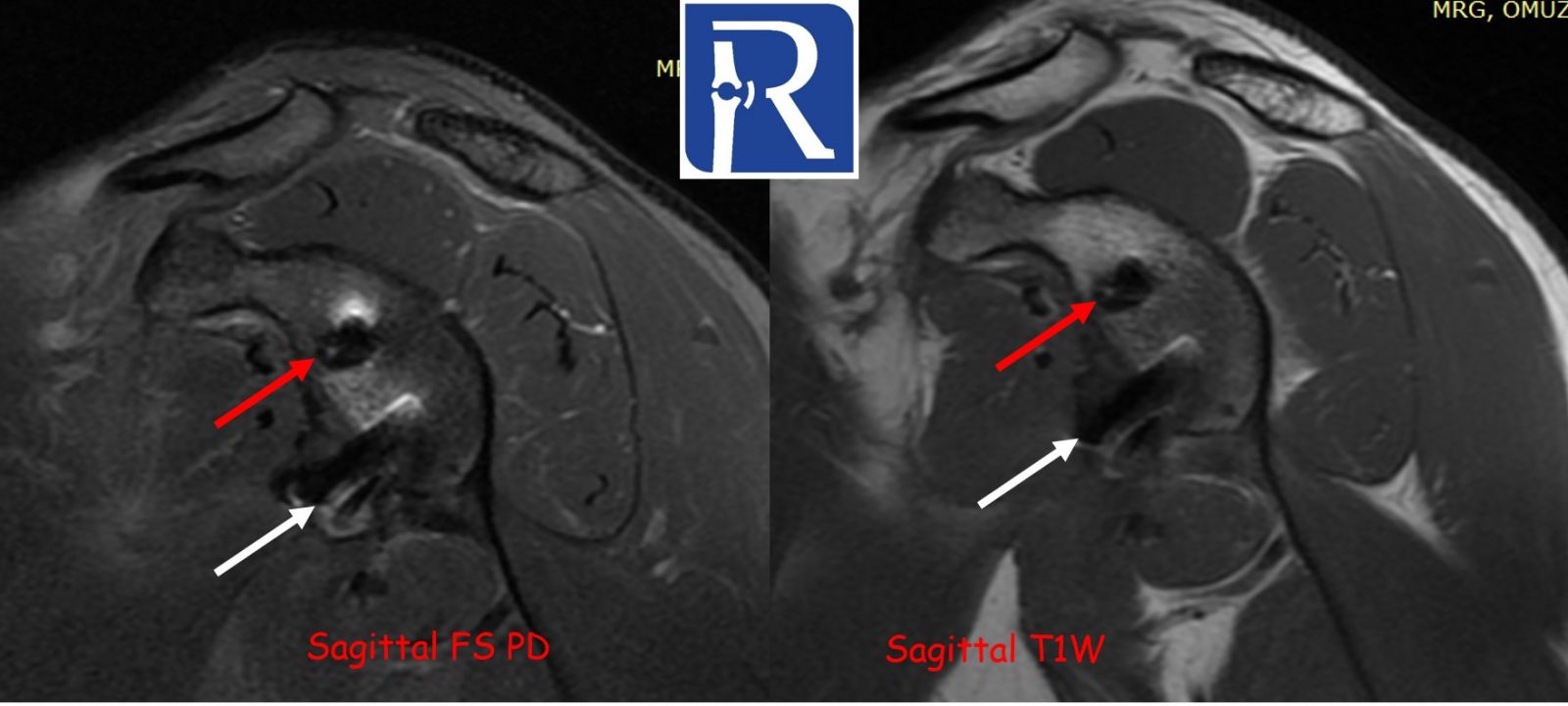
MRI Findings:
Sagittal plane images provide the most diagnostic information in shoulder evaluation and are particularly useful in identifying the type of prior surgical procedure.
- Susceptibility artifacts at the anteroinferior glenoid suggest a Bankart repair (white arrows). Corresponding metallic opacity on radiographs confirms the presence of a metallic anchor.
- Susceptibility artifacts at the superior glenoid support the presence of a labral repair (red arrows). This region appears radiolucent on radiographs, consistent with the presence of a bioabsorbable anchor.
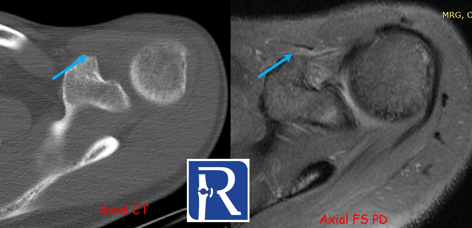
Additionally, marked edema and cortical irregularity are noted around the coracoid process.
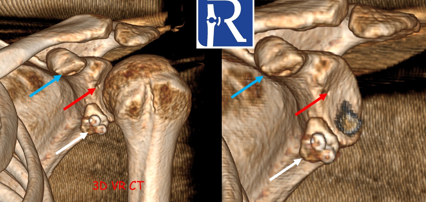
CT Findings:
CT demonstrates a bony defect at the coracoid process (blue arrows) and shows that the resected bone fragment has been fixed to the inferior glenoid defect with two anchors (white arrows). These findings are consistent with a Latarjet procedure.
A radiolucent anchor is also identified at the superior glenoid, corresponding to the site of the labral repair (red arrows).
Surgical and Radiologic Correlation:
In patients with recurrent shoulder dislocation and a significant Bankart defect, the Latarjet procedure is the preferred surgical option.
- Ideally, the transferred coracoid bone graft should be flush with the glenoid articular surface.
- A step-off of up to 2 mm is acceptable.
- A medial step-off >2 mm increases the risk of recurrent dislocation, whereas a lateral step-off raises the risk of impingement.
The Latarjet procedure involves transferring and fixing the coracoid bone block to the anteroinferior glenoid rim, typically using one or more screws, to augment the glenoid and restore anterior stability. It may be performed using open or arthroscopic techniques.
Radiologic Assessment Note:
For evaluation of step-off and graft positioning, CT and axial plane images are most useful. In contrast, sagittal plane MRI is the most informative for identifying the type of surgical procedure.
Sagittal images are indispensable for assessing impingement and adhesive capsulitis, as well as for determining the type and positioning of glenoid repair.
.jpeg)

0 COMMENTS
These issues are no comments yet. Write the first comment...