Sacroiliitis on MRI: Imaging Features and Differential Diagnosis
Case 1
Clinical presentation:
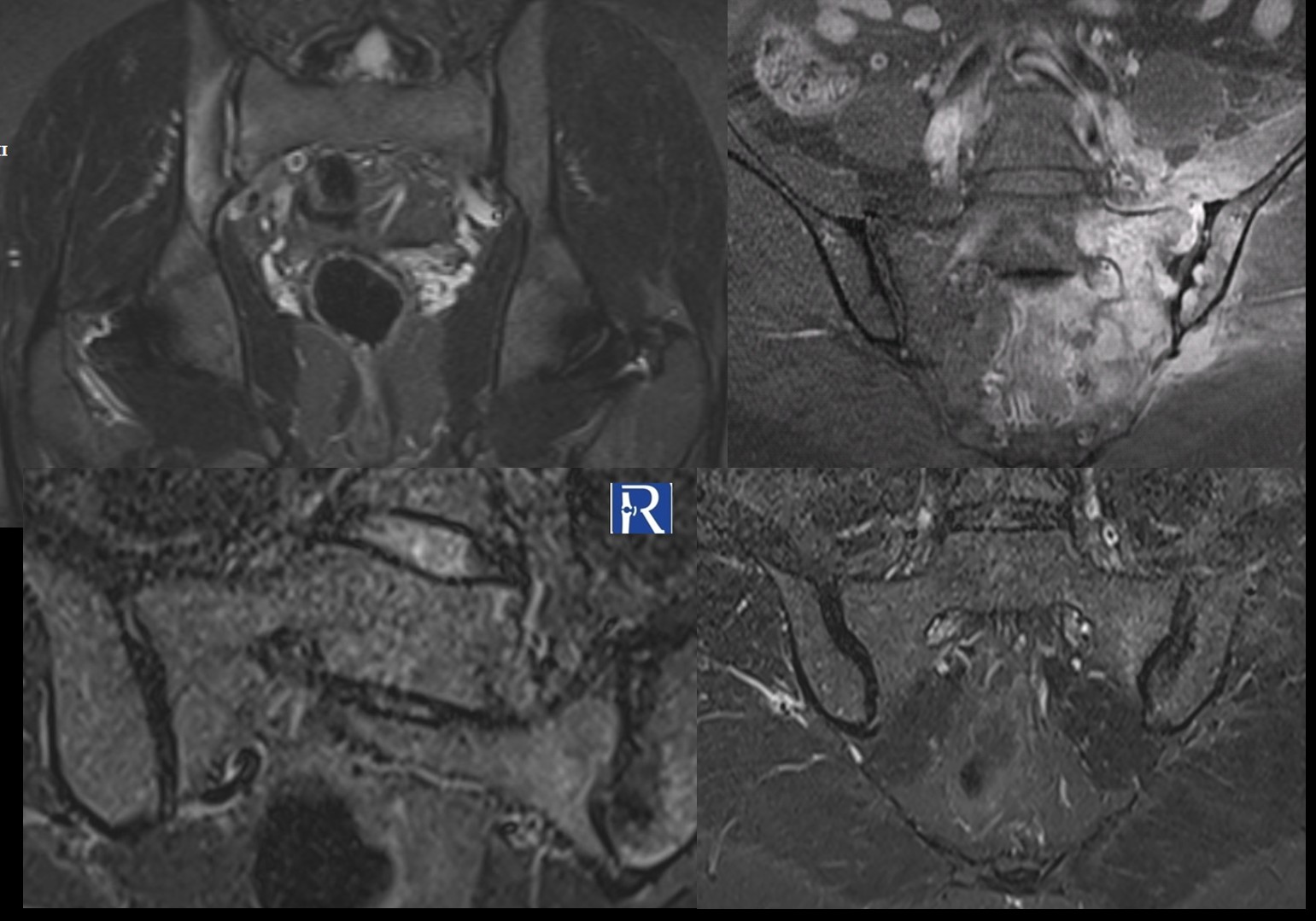
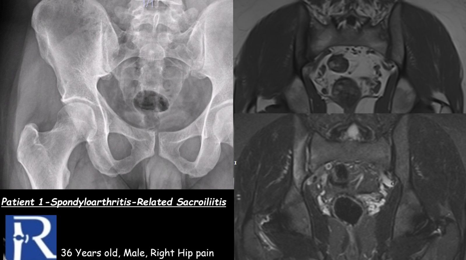
A 36-year-old man presenting with right hip pain. The patient is HLA-B27 positive.
MRI findings:
MRI demonstrates bone marrow edema–like signal changes involving the right sacroiliac joint, with predominant involvement of the iliac side. The imaging appearance is consistent with spondyloarthritis-associated sacroiliitis.
It should be noted that these findings are based on images acquired as part of a hip MRI examination; therefore, the images do not represent a dedicated, optimized sacroiliac joint MRI protocol.
Radiography:
Conventional radiographs demonstrate grade II sacroiliitis.
Case 2
Clinical presentation:
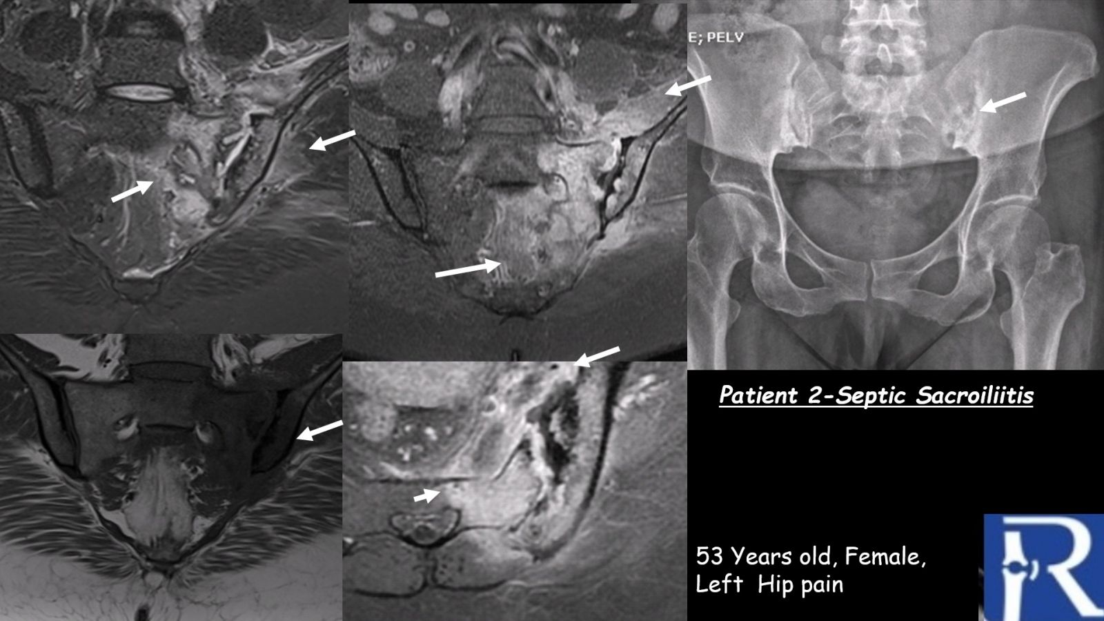
A 53-year-old woman presenting with left hip pain.
MRI findings:
MRI reveals extensive bone marrow edema centered on the left sacroiliac joint, accompanied by adjacent soft tissue edema. These findings are consistent with septic sacroiliitis.
Radiography:
Conventional radiographs demonstrate grade III sacroiliitis.
Case 3
Clinical presentation:
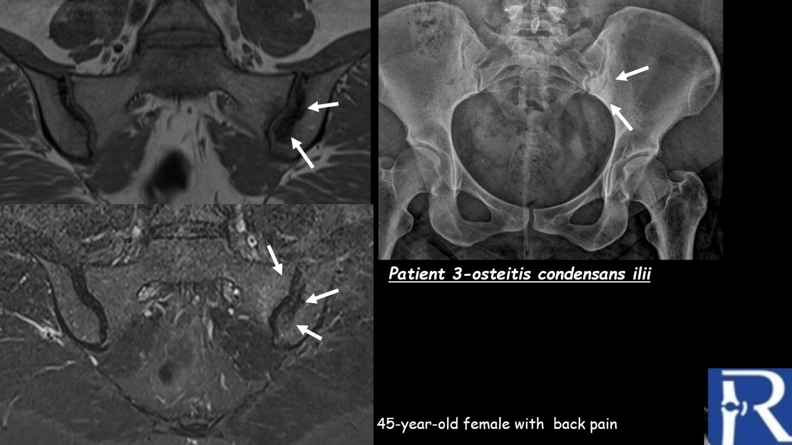
A 45-year-old woman presenting with back pain.
MRI findings:
MRI shows triangular-shaped periarticular sclerosis involving the left iliac bone, associated with mild bone marrow edema in the adjacent iliac and sacral bones at the left sacroiliac joint. The imaging findings are characteristic of osteitis condensans ilii.
Radiography:
Plain radiographs demonstrate triangular-shaped periarticular sclerosis of the left iliac bone.
Teaching Point
Sacroiliitis encompasses a broad differential diagnosis. Careful evaluation of the distribution of bone marrow edema, associated soft tissue changes, pattern of sclerosis, and clinical context is essential to differentiate inflammatory, infectious, and mechanical etiologies. In addition, interpretation of sacroiliac joint findings should take into account whether the images were obtained using a dedicated sacroiliac joint MRI protocol or incidentally visualized on hip MRI examinations.

0 COMMENTS
These issues are no comments yet. Write the first comment...