Ankle
Calcaneonavicular coalition
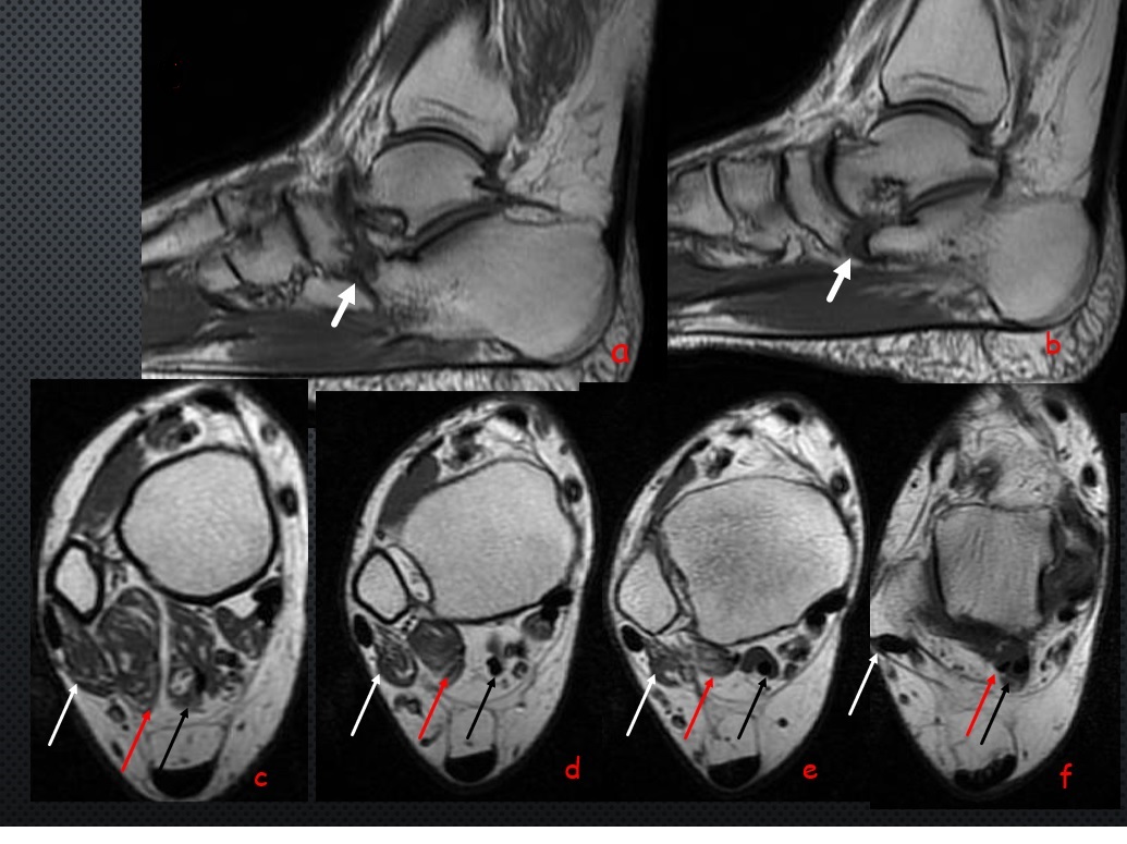
Figure Legends: Sagittal T1W images (a-b) demonstrates non osseous calcaneal-navicular coalition (white thick arrow) . Axial plane ( c-f) T1W images demonstrates accessory Peroneocalcaneus internus muscle (red arrow) which is located between peroneal (white arrow) and flexor hallucis longus (black arrow) tendons
Calcaneonavicular coalition is one of the two most common subtypes of the tarsal coalition. The other one is talocalcaneal coalition.
Multiple accessory muscles of ankle have been described in literature. Ankle accessory muscles are typically asymptomatic but may cause compressive neuropathy.
Various anatomical variations including accessory muscles-bones, coalitions, sesamoid bones can be found in ankle. These anatomical variations may cause ankle and foot problems.
MRI is helpful in assessment and characterization of coalition and accessory muscle and assessment of various ankle pathologies
In my experience T1Wsequences are more helpful for assessment and characterization of ankle anatomical variations. Sagittal planes are better for determination of the calcaneonavicular coalition. Axial planes are better for determination of the accessory muscles.

0 COMMENTS
These issues are no comments yet. Write the first comment...