Demographical and clinical details: 9 years old male child, admitted with left thigh pain
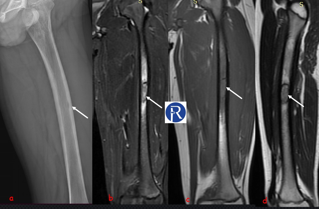
Image Details: Oblique-lateral femur X-Ray (a) shows the endosteal scalloping (white arrow) in anterior cortex of mid diaphyseal region, that is finding of Lodvick type 1 b (geographic)osteolytic lesion. Coronal STIR (b), T1W (b) and Sagittal T2W (d) images shows the heterogeneous low signal intensity tumoral lesion (white arrow) with accompanying perilesional edema-like signal changes. Bone biopsy was performed within the lesion due to aggressive appearance. The pathological diagnosis was consistent with eosinophilic granuloma (EG).
EG is benign proliferation of Langerhans cells usually accompanied with eosinophils, lymphocytes, neutrophils and scattered plasma cells. EG is monostotic in most cases(50-75% of cases). Monostotic form of Langerhans cell histiocytosis was classified as locally aggressive Hematopoietic bone tumor according to WHO classification (2020, 5th edition), whereas polyostotic form was classified as malignant hematopoietic bone tumor.
In my experience, Direct radiographs which are very important for the diagnosis of bone tumor should be carefully evaluated. Endosteal scalloping may be sign of serious bone tumor. EG, Ewing tumor and osteomyelitis should be considered in differential diagnosis when a bone tumor with peritumoral edema is detected in childhood. The signal intensity of peritumoral edema seen in EG is less than that seen in Ewing tumor and Osteomyelitis.

0 COMMENTS
These issues are no comments yet. Write the first comment...