Extramedullary Hematopoiesis in a patient with β-thalassemia major
Demographic and Clinical Details:A 50-year-old female with a known history of β-thalassemia major was admitted.
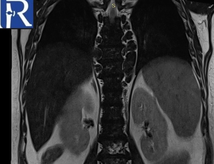
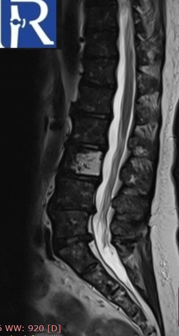
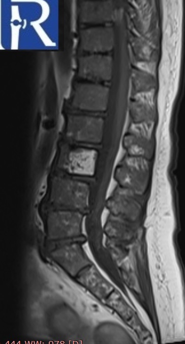
Image Details: Coronal and sagittal plane T1 and T2-weighted images reveal a diffuse decrease in signal intensity across all vertebral bodies, likely indicative of significant iron deposition resulting from repeated blood transfusions. Additionally, there are several paravertebral multilobulated soft tissue masses (indicated by white arrows) displaying markedly low signal intensity. The observed low signal intensity on T2-weighted images suggests a diagnosis of extramedullary hematopoiesis. A hemangioma is also noted in the L3 vertebra (black arrow). Furthermore, a diffuse decrease in signal intensity of the liver is evident, attributed to massive iron deposition (green arrow).

0 COMMENTS
These issues are no comments yet. Write the first comment...