Geyser Sign
Demographic and clinical details: 73 years, Female, pain and soft tissue mass in right shoulder
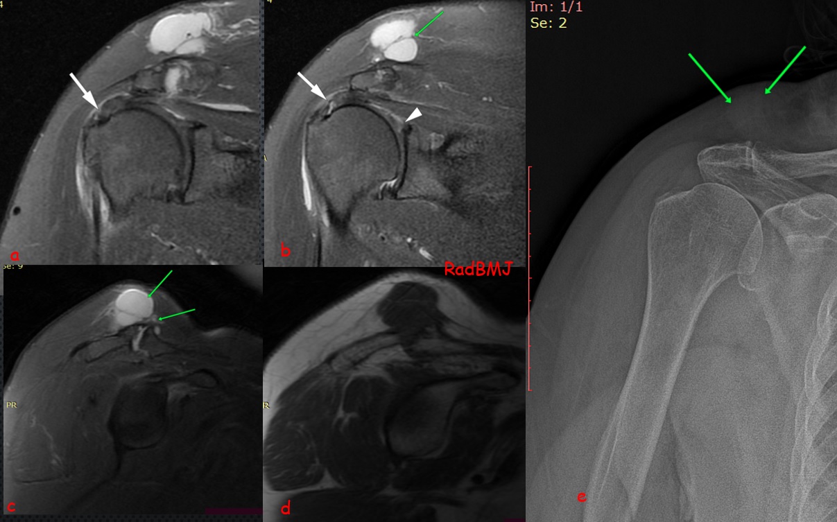
Image Details: Coronal (a,b), Sagittal (c) fat suppressed proton density and sagittal T1W(d) images demonstrate acromioclavicular (AC)joint effusion and cyst like lesion ( green arrows) located superior to the AC joint which is consistent with subcutaneous pseudotumor due to MRI Geyser sign. There is also bursal surface-ntratendinous partial thickness tear (white arrows) with associated superior labrum tear (white arrowhead). Plane Radiography shows the soft tissue opacity in subcutaneous tissue
AC joint cysts are rare presentation of shoulder pathologies. Most of the cysts were secondary to a chronic rotator cuff tear. Synovial fluid leaks through the AC joint to form a supraclavicular collection. This leakage when seen on a conventional arthrogram was seen “erupting” as a “Geyser” and hence was called the Geyser Sign.
In my experience, Demonstration of fluid like signal intensity within AC joint is important for the determination of MR geyser sign. When you see the geyser sign, you should look for the associated rotator cuff tear.

0 COMMENTS
These issues are no comments yet. Write the first comment...