MR Features of Dermatomyositis with Pulmonary Involvement
Case:
A 54-year-old male with a history of routine follow-up for interstitial pneumonia presented with a rash on the trunk and thighs, accompanied by significant lower extremity weakness. Histopathological evaluation of the rash confirmed a diagnosis of dermatitis.
MRI Findings:
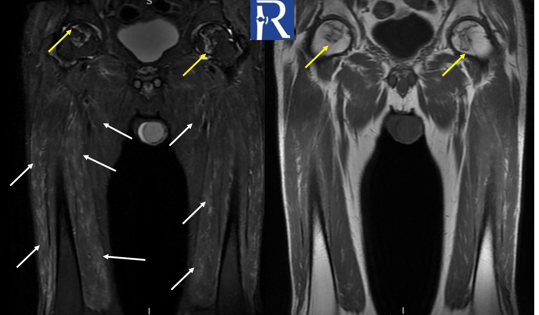
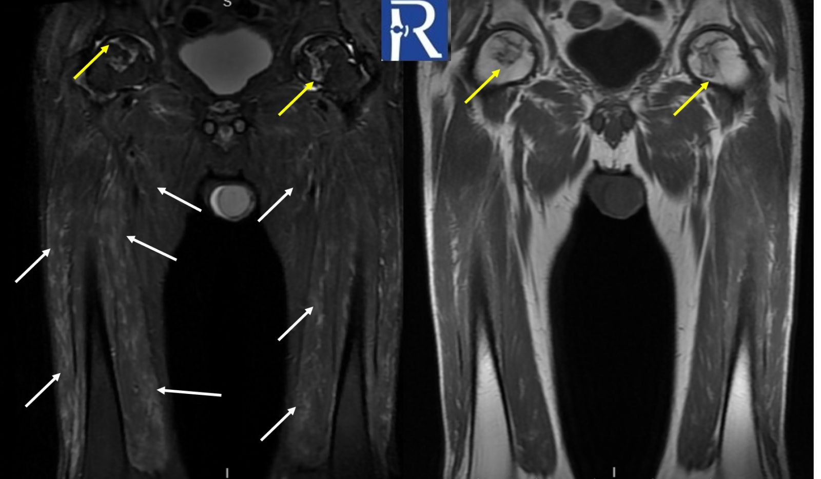
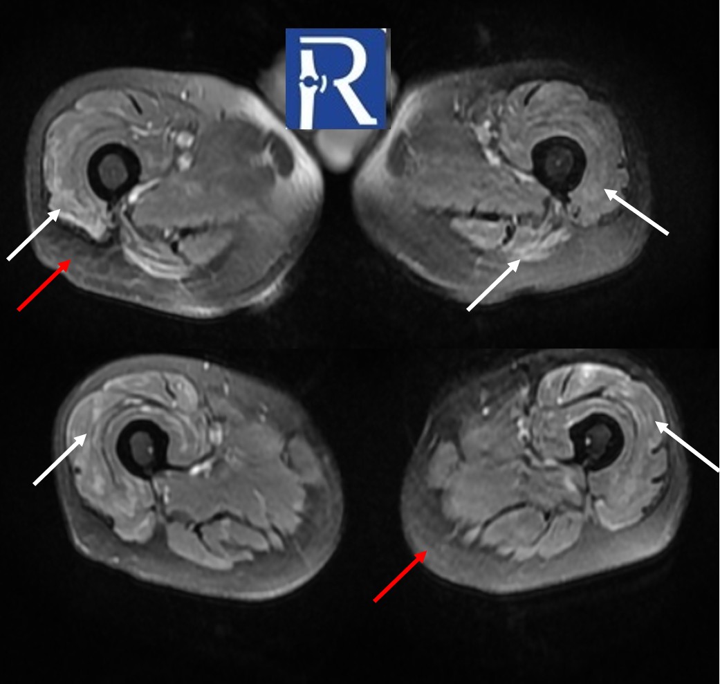
MRI of the both thigh reveals bilateral crescent-shaped, geographic lesions in the femoral heads consistent with osteonecrosis (formerly avascular necrosis) (yellow arrows). Additionally, there are bilateral, symmetrical, heterogeneous hyperintense signal changes—resembling edema—predominantly affecting the anterior muscle groups of the thighs (white arrows), indicative of myositis. There is also diffuse loss of subcutaneous fat tissue with focal areas of mildly increased T2 signal (red arrow).
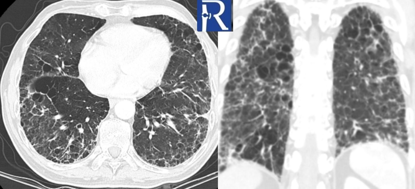
Thoracic CT demonstrates widespread subpleural thickening, reticular opacities, and a mild honeycombing pattern in both lungs.
Inflammatory myopathies frequently coexist with interstitial lung disease (ILD), and in some cases, ILD may dominate the clinical picture. A strong association has been reported between myositis-specific antibody (MSA) positivity and pulmonary involvement. The presence of ILD in patients with myositis is associated with increased morbidity and mortality

0 COMMENTS
These issues are no comments yet. Write the first comment...