Bone Marrow Involvement in Sarcoidosis
Case Presentation
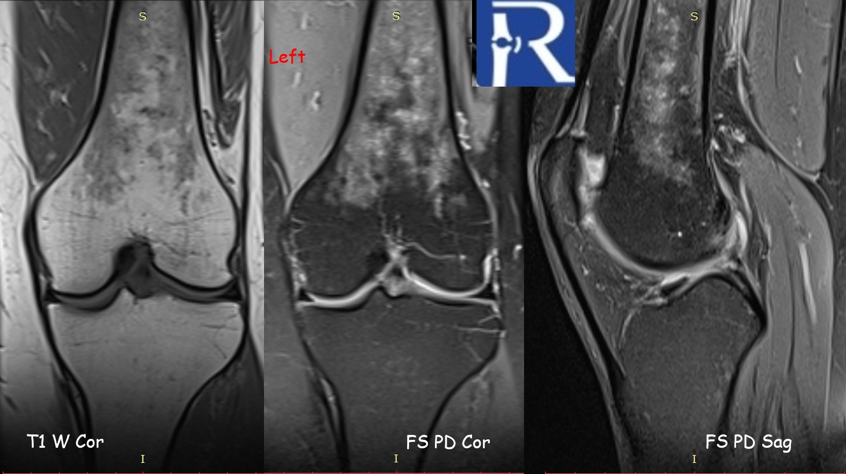
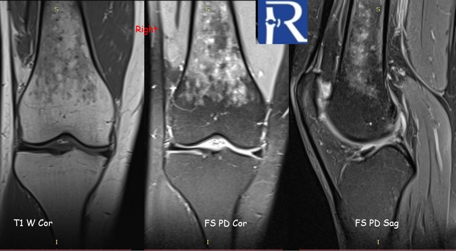
A 40-year-old female, who was under follow-up for sarcoidosis with known abdominal lymphadenopathy, presented with bilateral knee pain of insidious onset.
Initial plain radiographs of both knees were unremarkable. However, further evaluation with magnetic resonance imaging (MRI) revealed subcubic lesions infiltrating the bone marrow of both femurs, demonstrated on T1-weighted and fat-suppressed proton density sequences. Given the patient’s prior diagnosis of sarcoidosis, the imaging findings were interpreted as consistent with osseous involvement of the disease.
Sarcoidosis is a chronic multisystem granulomatous disorder characterized histologically by the presence of non-caseating granulomas. The lungs are involved in over 90% of cases, but extrapulmonary manifestations, including those affecting the skin, lymph nodes, and eyes, are also common. Skeletal sarcoidosis, however, is a rare manifestation, more frequently affecting the small bones of the hands and feet, with involvement of long bones being less commonly reported.
MRI is the most sensitive imaging modality for detecting skeletal sarcoidosis, particularly for evaluating changes in the bone marrow. However, the imaging appearance is nonspecific and may mimic other pathologies such as metastatic disease, lymphoma, multiple myeloma, or osteomyelitis. Correlation with clinical history and, when necessary, histopathologic confirmation is essential for accurate diagnosis.
.jpeg)

0 COMMENTS
These issues are no comments yet. Write the first comment...