Seeing Beyond the Spine: Coronal STIR Reveals a Hidden Plexiform Neurofibroma

Abstract:
Plexiform neurofibroma is a rare benign tumor of the peripheral nerves, often associated with Neurofibromatosis Type 1 (NF1). We present the case of a 60-year-old male with low back pain whose routine lumbar MRI appeared unremarkable on sagittal and axial planes. However, the coronal STIR sequence incidentally revealed a 22 × 2.5 cm grape-like, heterogeneously enhancing soft-tissue mass extending from the greater sciatic foramen along the left sciatic nerve. The lesion’s morphology and signal characteristics were consistent with a plexiform neurofibroma. This case underscores the importance of including fluid-sensitive coronal sequences in standard spinal MRI protocols for detecting paraspinal and extraspinal pathologies that may otherwise remain occult.
Clinical Information
A 60-year-old male patient underwent lumbar MRI for evaluation of chronic low back pain. The patient had no known history or clinical features of Neurofibromatosis Type 1 (NF1).
Imaging Findings
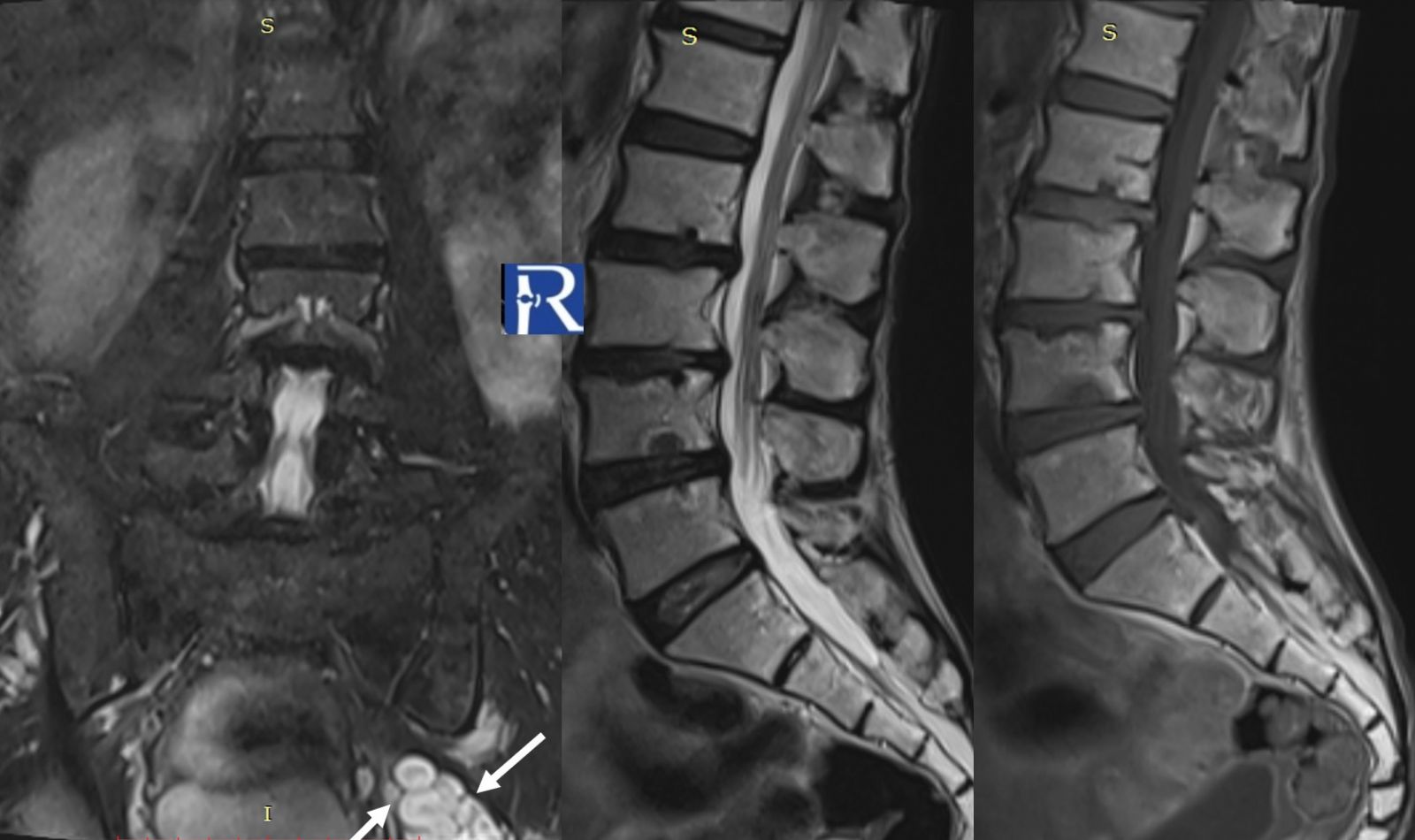
A routine lumbar MRI revealed findings consistent with lumbar spondylosis and discopathy. However, on the coronal STIR sequence, a grape-like soft tissue mass with an internal target sign was incidentally detected adjacent to the left piriformis muscle.
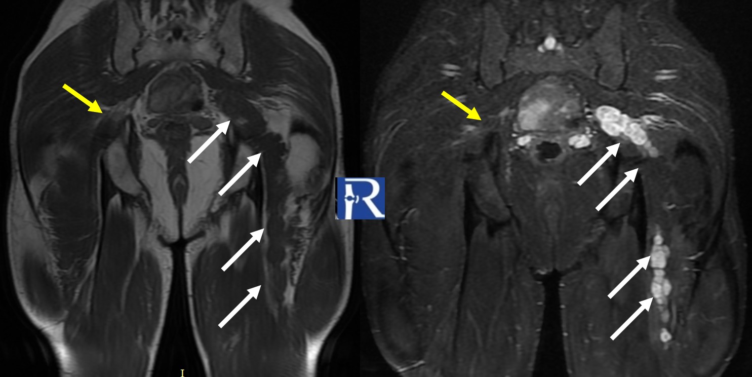
Subsequent hip MRI revealed a grape-like, heterogeneously enhancing soft tissue lesion extending from the greater sciatic foramen inferiorly along the course of the left sciatic nerve.
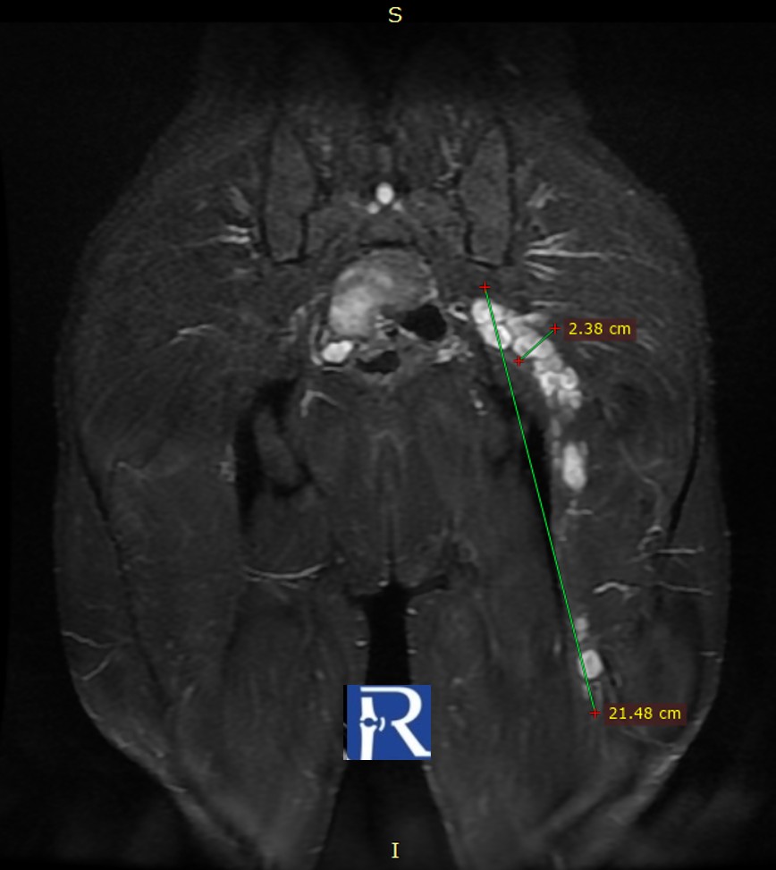
The lesion measured approximately 22 cm craniocaudally and 2.5 cm transversely, showing heterogeneous enhancement following contrast administration.
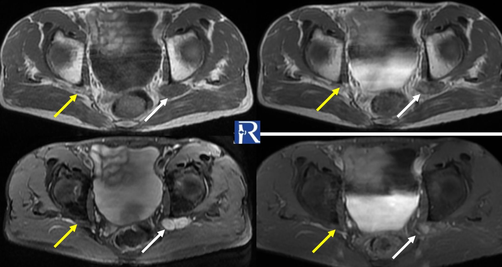
White arrows indicated the left sciatic nerve, and yellow arrows the right sciatic nerve for comparison.
Diagnosis
Plexiform Neurofibroma of the Sciatic Nerve
Discussion
Plexiform neurofibroma is an uncommon benign tumor of peripheral nerves (WHO Grade I), representing a diffuse proliferation of all neural elements. It typically occurs in association with Neurofibromatosis Type 1 and is considered pathognomonic for the disease.
However, as in this case, it may occasionally present sporadically, without any clinical signs of NF1.
Plexiform neurofibromas often display a “grape-like” or multilobulated configuration, following the course of the affected nerve. On MRI, they characteristically show multiple target signs on T2/STIR sequences — a central area of low signal intensity surrounded by hyperintense myxoid tissue — and heterogeneous enhancement after contrast administration.
Unlike localized neurofibromas, plexiform variants have a potential for malignant transformation into malignant peripheral nerve sheath tumors (MPNSTs), necessitating careful radiologic and clinical follow-up.
Teaching Point
This case highlights the importance of incorporating a fluid-sensitive coronal sequence, such as coronal STIR, into routine lumbar MRI protocols.
Although the primary indication was low back pain, the coronal sequence enabled the incidental detection of a clinically significant extraspinal lesion that would likely have been missed on sagittal and axial planes alone.
A comprehensive spinal MRI protocol should, therefore, incorporate coronal imaging, which enhances detection of paraspinal and extraspinal pathologies, particularly those extending through neuroforamina or along major peripheral nerves.

0 COMMENTS
These issues are no comments yet. Write the first comment...