Morton Neuroma
Demographical and Clinical details: 50 y, F, forefoot pain and numbness
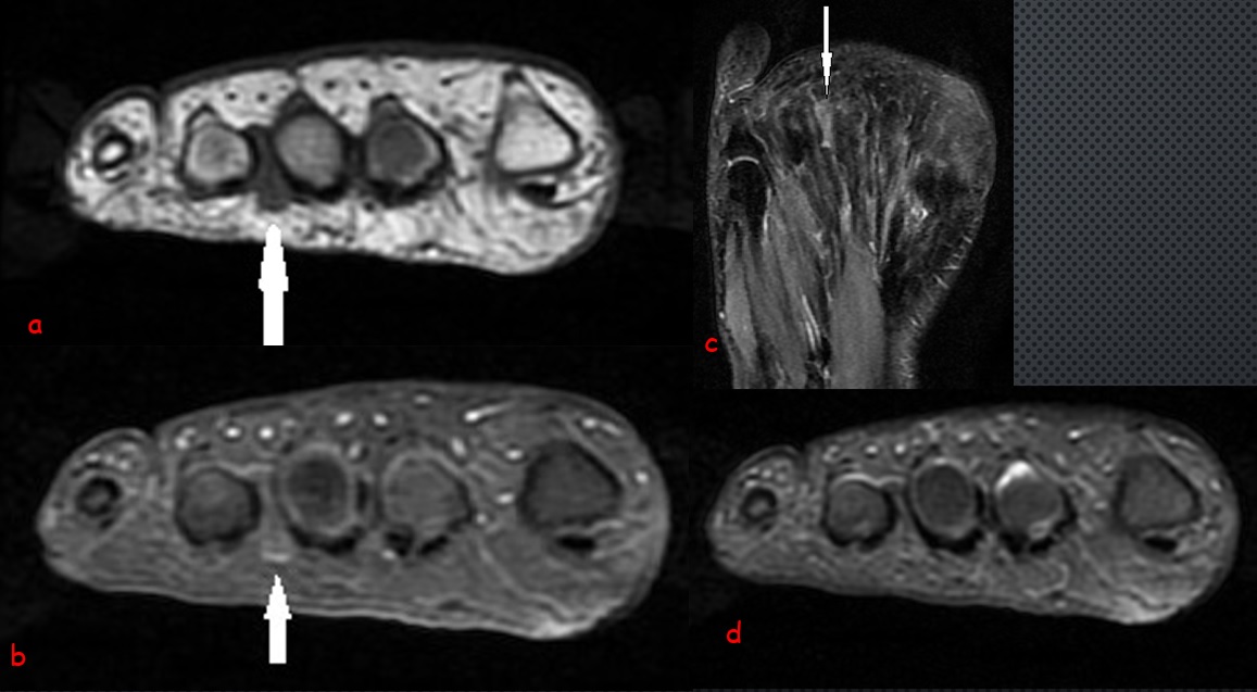
Radiological Findings: Coronal T1-weighted image(a) of right foot shows the low-signal-intensity dumbbell-shaped nodule at third intermetatarsal space (arrow), with its epicenter plantar to deep transverse intermetatarsal ligament. Coronal contrast enhanced fat saturated T1 W image(b) shows the slightly heterogenous enhancement which indicates the solid nature of lesion. Axial and coronal fat saturated PD images (c, d) slightly hypointensity of the lesion which excludes the diagnosis of intermetatarsal bursitis. Radiological Findings is consistent with Morton neuroma.
Morton neuromas are not true tumor, rather they re focal areas of symptomatic perineural fibrosis around a plantar digital nerve of foot. T They re most commonly seen between 3rd and 4th metatarsal heads, but less commonly seen between 2nd and 3rd metatarsal heads.
In my expertise, coronal plane T1 weighted images are better for the determination of Morton neuroma.It should be differentiated from intermetarsal bursitis.

0 COMMENTS
These issues are no comments yet. Write the first comment...