Hallux sesamoid Fracture
Demographic and clinical details: 53 years old female, admitted with right foot pain
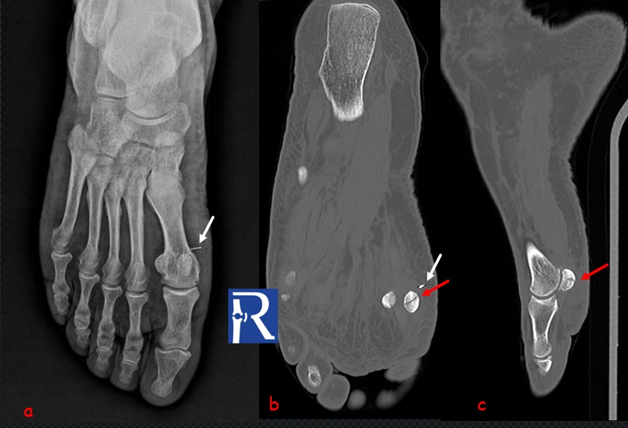
Image Details; AP foot radiograph shows the small metallic density in soft tissue medial to the medial hallux sesamoid, consistent with metallic foreign body (white arrows). (a)Non-displaced fracture is seen in medial sesamoid on axial (b) and sagittal(c) CT images (red arrows).
Fractures can be missed on direct radiography if they are non-displaced and located in complex anatomical parts. CT is more sensitive and specific than plain radiographs for the assesment of hallux sesamoid fractures
Hallux sesamoids are prone to weight bearing stress injury, with the medial sesamoid fractured more frequently than the lateral one
It must be differentiated from the bipartite sesamoid bone. Well corticated margins of bipartite bone fragments are helpful sign for differentiation

0 COMMENTS
These issues are no comments yet. Write the first comment...