Are We Missing Plantar Vein Thrombosis?

Case Presentation
Plantar vein thrombosis (PVT) has traditionally been described as a rare cause of foot pain. However, increasing awareness and more frequent use of high-resolution MRI may be revealing that this entity is underrecognized rather than truly uncommon.
We report a 60-year-old woman with rheumatoid arthritis who presented with persistent lateral plantar foot pain refractory to NSAIDs and injection therapy. There was no definite recent trauma.
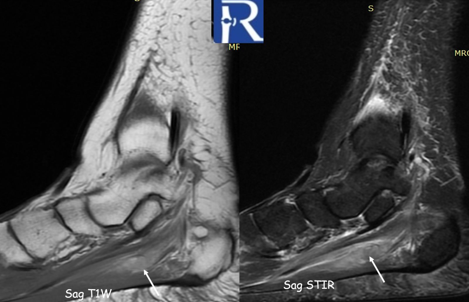
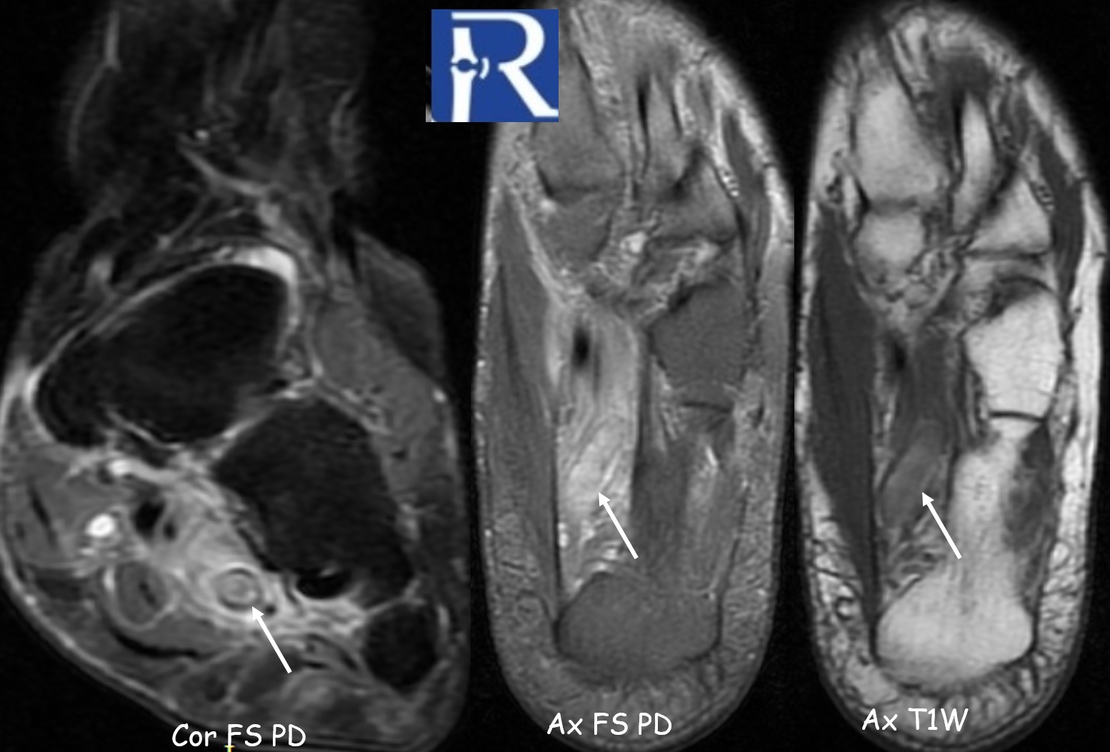
MRI of the left ankle demonstrated:
- Loss of normal venous flow void in the lateral plantar vein
- Venous dilatation
- T1 hyperintense intraluminal signal compatible with subacute thrombus
- Hyperintense thrombus on proton density fat-suppressed sequences
- Prominent perivascular soft tissue edema extending into adjacent musculature
Perivascular edema was the most conspicuous indirect sign.
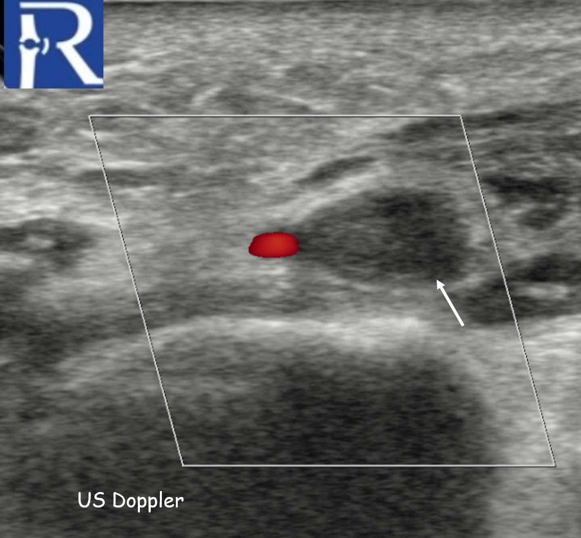
The case was initially reported as negative for venous thrombosis on routine Doppler examination. However, when reassessment was specifically requested with attention to the plantar venous system, duplex ultrasound confirmed lateral plantar vein thrombosis
Discussion
Plantar vein thrombosis is sparsely reported in the literature and remains a relatively unfamiliar diagnosis in daily clinical practice. Several case reports and small series suggest that patients typically present with nonspecific plantar or lateral foot pain, often mimicking musculoskeletal disorders such as plantar fasciitis or tendinopathy.
A critical issue appears to be protocol-related: plantar veins are not routinely included in standard lower extremity DVT ultrasound examinations. As a consequence, thrombosis confined to these distal venous structures may go undetected unless specifically targeted.
MRI findings described in the literature include:
- Loss of normal venous flow void
- Venous enlargement
- Intraluminal signal abnormality depending on thrombus age
- Perivascular edema
Perivascular edema, in particular, may serve as an important indirect imaging clue prompting focused vascular assessment.
Our increasing identification of plantar vein thrombosis on MRI, combined with initially negative routine Doppler studies, supports the hypothesis that this condition may be underdiagnosed rather than rare.
Clinical and Imaging Implications
When venous thrombosis is clinically suspected and patients present with:
- Persistent unexplained lateral or plantar foot pain
- Inflammatory or hypercoagulable risk factors
- Negative initial Doppler findings
Targeted inclusion of the medial and lateral plantar veins in duplex ultrasound evaluation may be appropriate.
Systematic awareness and protocol adjustment in selected cases could improve diagnostic accuracy without substantially increasing examination time.
Learning Points
• Plantar vein thrombosis may be underrecognized due to omission from routine DVT protocols.
• MRI is particularly valuable when Doppler findings are negative but suspicion persists.
• Perivascular edema is an important indirect imaging sign.
• Focused reassessment of the plantar venous system can change management.
If confirmed in larger series, these observations may justify reconsideration of distal venous assessment strategies in patients with persistent unexplained plantar foot pain.

0 COMMENTS
These issues are no comments yet. Write the first comment...