Chalk-Stick Fracture in Ankylosing Spondylitis

Clinical Background
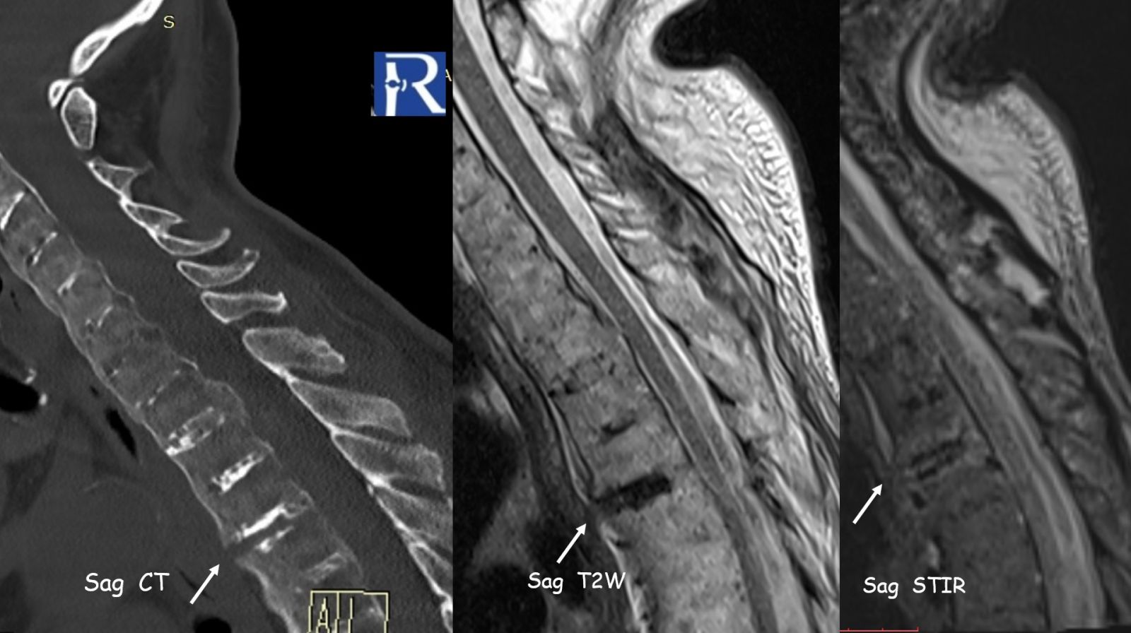
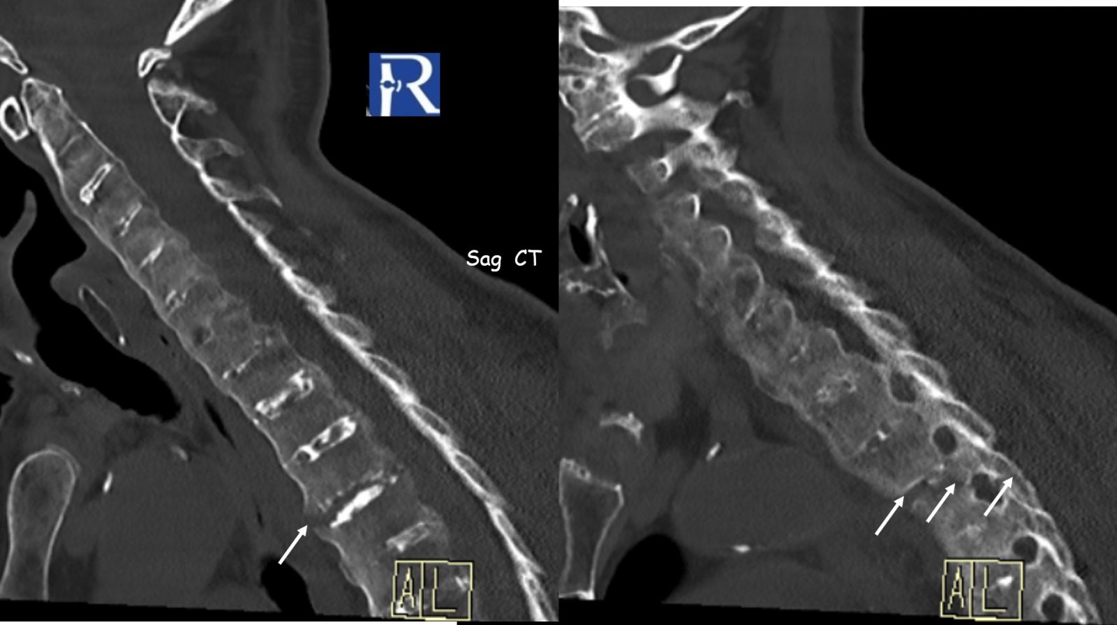
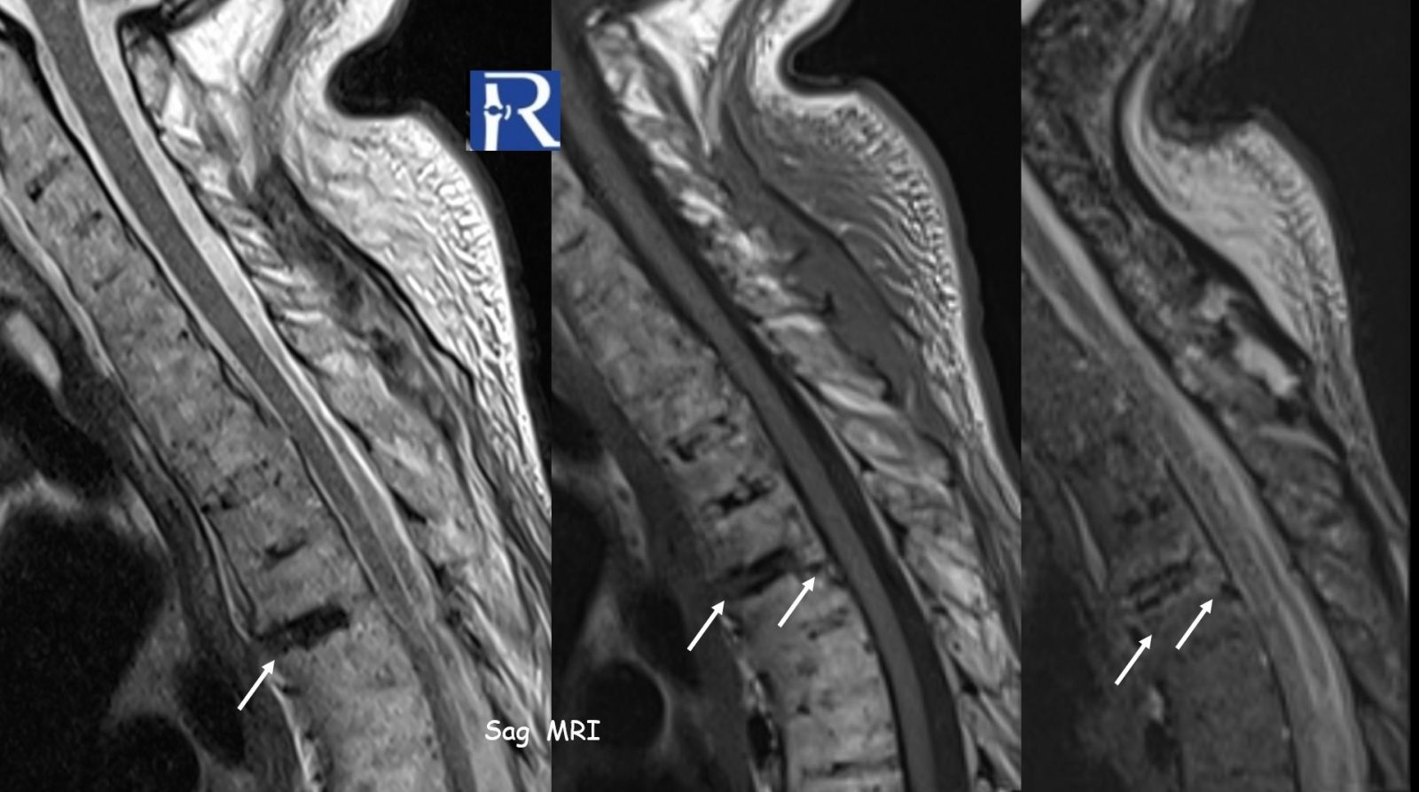
A 77-year-old patient with Ankylosing Spondylitis presented after a low-energy fall with acute thoracic pain.
Imaging Findings
- Extensive bridging syndesmophytes throughout the spine
- Complete spinal ankylosis (“bamboo spine” morphology)
- At the T3–T4 level, a transdiscal fracture is observed
- Fracture line extends:
- From anterior fused syndesmophyte
- Through the intervertebral disc
- Into the posterior elements (facet joints)
- Associated marrow edema and soft tissue changes on MRI
This represents a classic “chalk-stick fracture”, a transverse fracture through a rigid, ankylosed spine.
Diagnosis
Chalk-stick fracture of the ankylosed thoracic spine
Pathophysiology (Why it happens?)
- Ankylosed spine behaves as a long rigid lever arm
- Loss of segmental mobility > stress concentration at weak points
- The intervertebral disc space becomes the weakest link
- Even minor trauma can result in highly unstable three-column fractures
Key Radiological Insight
In ankylosed spines, the fracture is not where you expect—it is where the spine is weakest: the disc level
TEACHING PEARLS
1. Always suspect fracture—even after trivial trauma
- Ankylosed spine = “fracture-prone spine”
- Low-energy trauma can cause catastrophic instability
2. The fracture is typically transdiscal
- Most occur at disco-vertebral junctions
- Especially:
- Lower cervical
- Cervicothoracic junction
- Thoracolumbar junction
3. Look beyond plain radiographs
- X-ray may miss fractures
- CT (especially sagittal reconstructions) is essential
- MRI → evaluates:
- Bone marrow edema
- Ligamentous injury
- Spinal cord involvement
4. Follow the syndesmophytes
- Continuity disruption = fracture
- Carefully track the anterior longitudinal ossification line
5. Think “three-column injury”
- These fractures are usually:
- Highly unstable
- Extend through anterior + middle + posterior columns
- High risk of neurological deterioration
6. Don’t forget the mimickers / associations
Also seen in:
- Diffuse Idiopathic Skeletal Hyperostosis
- Surgical spinal fusion
- Ossified ligaments (OPLL / OLF)
“An ankylosed spine is not strong—it is brittle.”
“In trauma, systematically check every disc level.”
“Missed fracture = delayed neurological catastrophe.”

0 COMMENTS
These issues are no comments yet. Write the first comment...