Liposclerosing Myxofibrous Tumor; Typical Location, Classical Radiologic Features
Clinical Findings: Incidental finding in a 42-year-old after trauma. Typical location, classic radiologic features
Imaging Findings:
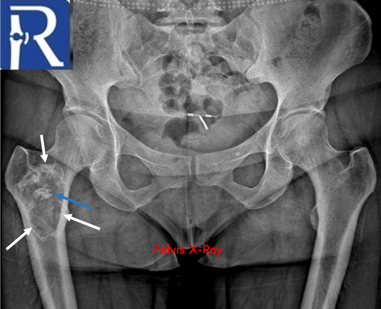
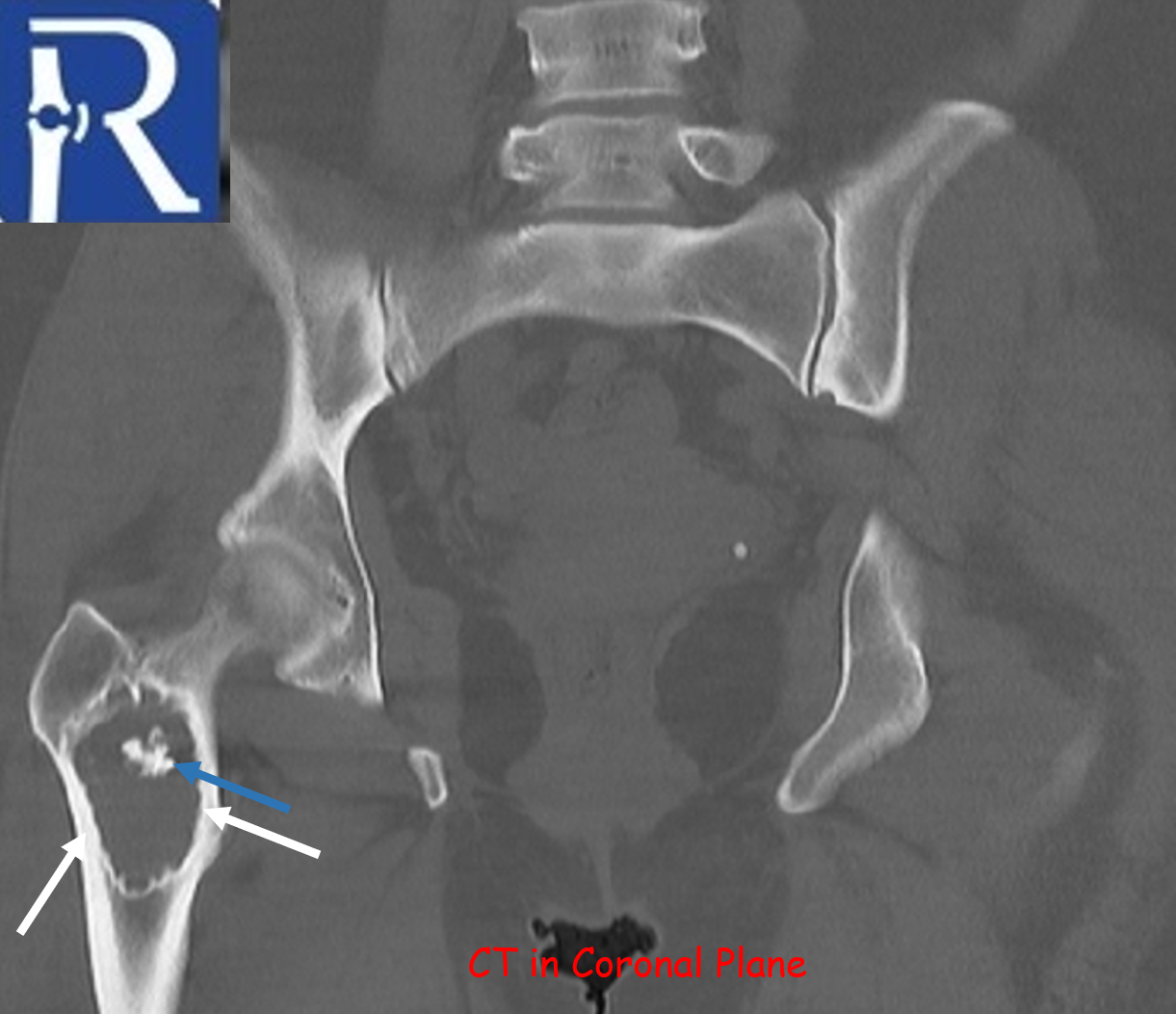
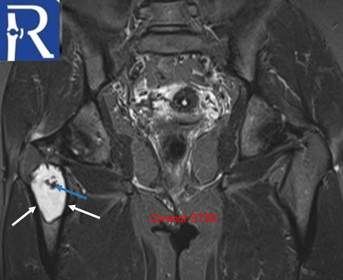
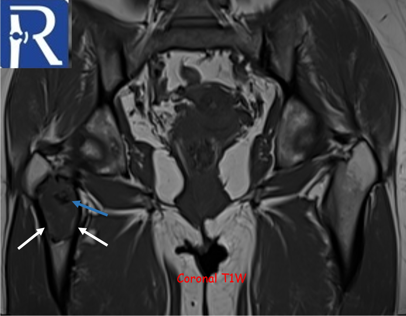
A geographic lytic lesion of Type I A pattern (white arrows) is observed on the right femur intertrochanteric-subtrochanteric area on the pelvic radiograph. The lesion is well-circumscribed and has a narrow transition zone. The internal content contains calcification (indicated by the blue arrow). Periosteal reaction is not visualised. The lesion demonstrates sclerotic areas corresponding to internal calcified foci. The lesion is compatible with liposclerosing myxofibrous tumors. In CT, the lesion matrix structures are more clearly visible, and the cortex appears intact. Calcification areas in the internal structure are more clearly observed. In MRI, the lesion appears as a low signal on the T1-weighted (T1W) sequence and as a high signal on the short tau inversion recovery (STIR) sequence, representing the myxoid content. Additionally, the calcification areas within the lesion exhibit low signals in both sequences.
Liposclerosing myxofibrous tumors (LFTs) are known as benign fibro-osseous lesions with a predilection for the femoral intertrochanteric region. They typically affect adults in the fourth decade of life. They exhibit distinctive radiological data and various histopathological findings, with a particular prevalence of malignant transformation. In the latest WHO Bone and Soft Tissue Tumors (2020), LSMFTs do not have their own section and are only mentioned in the section on fibrous dysplasia.

0 COMMENTS
These issues are no comments yet. Write the first comment...