Solid-Appearing Hand Mass Mimicking a Ganglion Cyst: MRI Features of Monophasic Fibrous Synovial Sarcoma

Clinical Presentation
A 45-year-old male presented with a slowly enlarging palpable mass on the volar aspect of the left hand. The patient reported mild discomfort during gripping activities but denied trauma, redness, or systemic symptoms. The initial clinical impression favored a ganglion cyst, and MRI was performed for further characterization.
MRI Findings
Non-contrast MRI
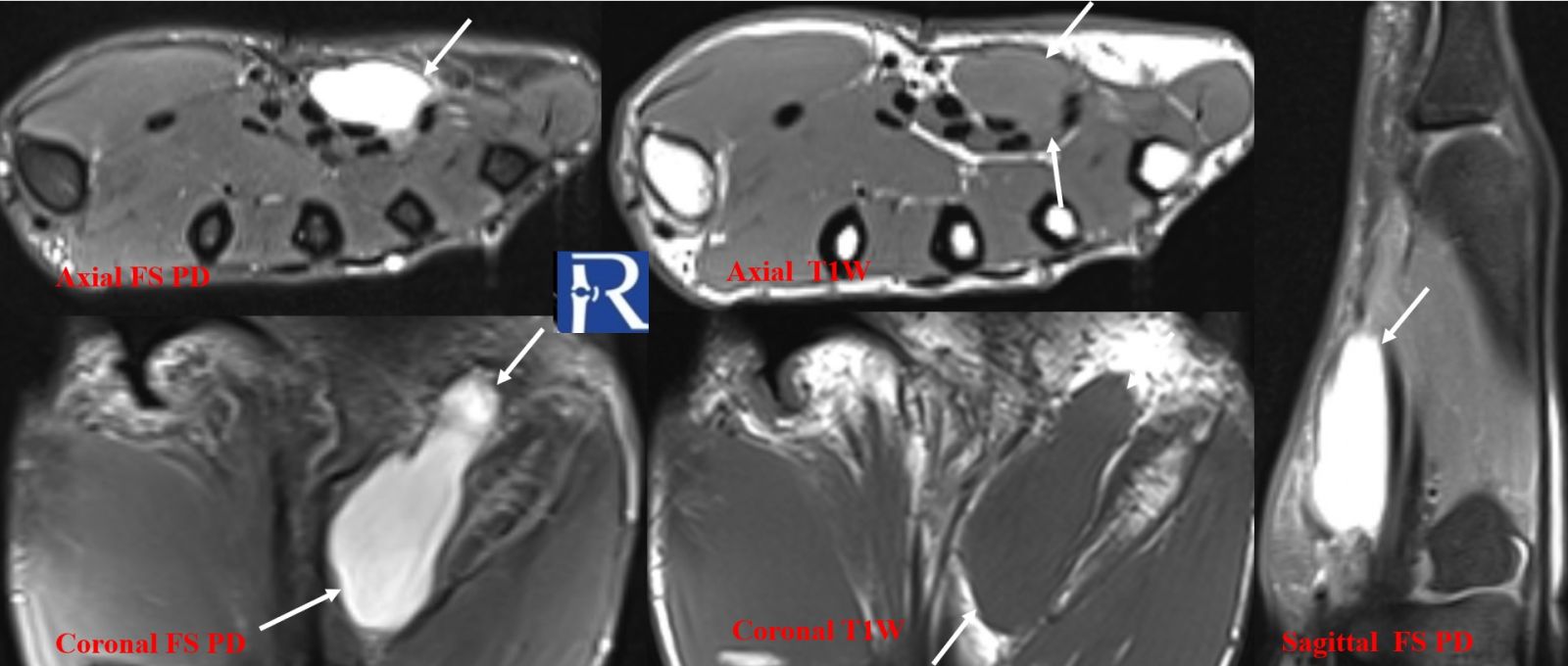
- T1-weighted images:
The lesion appeared isointense to adjacent muscle, an atypical feature for a simple ganglion cyst, which usually demonstrates low T1 signal. - Fluid-sensitive fat-suppressed sequences:
A well-defined soft-tissue mass was noted along the volar aspect of the 3rd, 4th, and 5th flexor tendons, mildly hyperintense relative to muscle (arrows).
The appearance initially suggested a cystic lesion.
Contrast-enhanced MRI
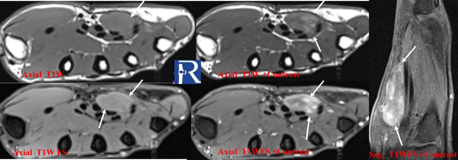
- Post-contrast T1 fat-suppressed sequences:
The lesion demonstrated heterogeneous internal enhancement, indicating solid tissue rather than a simple cyst.
No definitive fluid–fluid levels, necrotic areas, or macroscopic fat were identified.
Additional Features
- No evidence of bone erosion or cortical destruction.
- No significant perilesional edema.
- The mass displayed a subtle lobulated contour, a feature sometimes associated with soft-tissue sarcomas.
Histopathology
Ultrasound-guided core biopsy was performed.
Microscopic examination demonstrated uniform spindle cells arranged in fascicles, consistent with monophasic fibrous synovial sarcoma.
Discussion
Synovial sarcoma is an aggressive soft-tissue tumor that typically affects young adults and often arises near large joints of the extremities. Despite its name, it does not arise from synovium but from pluripotent mesenchymal cells.
In the hand, synovial sarcoma is rare yet clinically significant due to:
- frequent misdiagnosis as benign lesions (ganglion cyst, fibroma, GCT of tendon sheath),
- its slow growth pattern,
- and delays in appropriate referral and treatment.
MRI is the cornerstone of evaluation, and certain features should raise suspicion:
- Isointense to muscle on T1 (unusual for a cyst),
- Heterogeneous enhancement,
- Lobulated or infiltrative margins,
- Triple signal intensity on T2 (not always present).
In this case, the key diagnostic clue was the discrepancy between cyst-like appearance on fluid-sensitive sequences and unexpected isointensity on T1, prompting contrast administration, which revealed the solid nature of the lesion.
Teaching Points
- Not all “cyst-like” lesions are cysts; T1 isointensity to muscle should raise suspicion for a solid tumor.
- Contrast-enhanced MRI is essential for accurate differentiation between cystic and solid soft-tissue masses.
- Synovial sarcoma often mimics benign hand lesions and may lead to delayed diagnosis.

0 COMMENTS
These issues are no comments yet. Write the first comment...