Bone metastasis
Demographic and clinical details: 64-year-old, male patient, admitted with swelling and pain in the left arm-
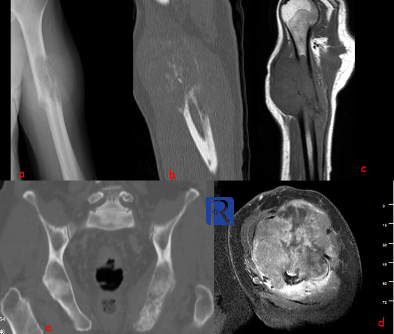
Image Details: Humerus frontal X-ray (a) shows the expansile -geographic osteolytic destructive lesion extending into the soft tissue with accompanying pathological fracture. Reconstructed CT image of sagittal plane shows the calcification within the lesion. Sagittal T1W and axial contrast enhanced fat suppressed T1W images shows the extension of lesion into the soft tissue and tumor necrosis. High grade chondrosarcoma can be considered among the differential diagnoses according to these radiological findings (aggressive malignant appearance and calcification within lesion), but There is one more slightly expansile lesion in left ischium which suggest the diagnosis of metastases. Pathological diagnosis is rectum cancer metastasis. Bone metastases are classified in the group of other mesenchymal tumors according to the WHO 2020 classification. Skeletal metastases account for 70% of all malignant bone tumor. The majority of metastases to bone are asymptomatic but some patients with bone metastasis may suffer from pain due to pathological fracture as occur in our patient.
The distribution of skeletal metastases generally reflects the distribution of red marrow.
In my experience, X-Ray is also important for the evaluation of soft tissue masses because X-ray shows that the soft tissue mass originates from bone.

0 COMMENTS
These issues are no comments yet. Write the first comment...